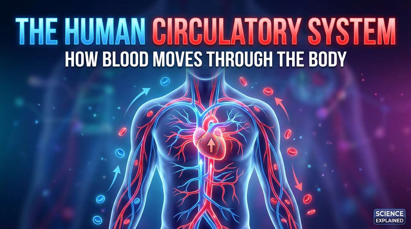
The Human Circulatory System: How Blood Moves Through the Body
Introduction
Right now, without any conscious effort on your part, your heart is contracting and relaxing roughly once every second, pushing blood through a network of vessels so extensive that, laid end to end, they would stretch far enough to circle the Earth more than twice. This happens continuously, from before you were born until the moment you die, without you ever needing to think about it.
The circulatory system — also called the cardiovascular system — is the body's transport network. It carries oxygen from the lungs to every living cell, removes carbon dioxide and waste products, distributes nutrients absorbed from digested food, transports hormones from glands to their target organs, and forms a crucial part of the body's defence against infection. Without it, no other system in the body could function for more than a few minutes.
This article explores the circulatory system in depth: its structures, how it works, the different types of circulation within the body, common disorders that can affect it, and how this topic is examined in WAEC, NECO, and JAMB Biology.
Why Living Things Need a Circulatory System
Every cell in the human body needs a continuous supply of oxygen and nutrients to carry out respiration and produce energy, and every cell must also get rid of waste products such as carbon dioxide. In very small organisms, materials can simply diffuse directly between cells and the environment, because no cell is very far from the outside.
The human body, however, is far too large and complex for diffusion alone to supply every cell in time. Some cells — such as those deep inside muscles or organs — are centimetres away from the nearest surface. Without a transport system, these cells would receive oxygen and nutrients far too slowly to survive. The circulatory system solves this problem by actively pumping blood throughout the body, ensuring that no cell is ever more than a very short distance from a capillary carrying fresh supplies.
The Main Components of the Circulatory System
The human circulatory system consists of three main components working together:
- The heart — a muscular pump that drives blood around the body.
- Blood vessels — a network of tubes (arteries, veins, and capillaries) through which blood travels.
- Blood — the fluid tissue that actually carries oxygen, nutrients, hormones, and waste products.
The Heart: Structure and Function
The heart is a hollow, muscular organ roughly the size of a closed fist, located slightly to the left of the centre of the chest, protected by the rib cage. It is made of a special type of muscle tissue called cardiac muscle, which is capable of contracting rhythmically and continuously throughout a person's entire life without becoming fatigued in the way skeletal muscle does.
The Four Chambers of the Heart
The heart is divided into four separate chambers, each with a specific role:
- Right atrium: Receives deoxygenated blood returning from the body.
- Right ventricle: Pumps deoxygenated blood to the lungs.
- Left atrium: Receives oxygenated blood returning from the lungs.
- Left ventricle: Pumps oxygenated blood out to the rest of the body.
The atria (upper chambers) are relatively thin-walled, since they only need to push blood a short distance into the ventricles below them. The ventricles (lower chambers) have much thicker, more muscular walls, because they must generate enough force to push blood either to the lungs or all the way around the entire body.
Notably, the wall of the left ventricle is significantly thicker than that of the right ventricle. This is because the left ventricle must generate enough pressure to pump blood through the systemic circulation — the entire body, from the brain to the toes — while the right ventricle only needs to pump blood the relatively short distance to the nearby lungs.
The Heart Valves
Blood must flow through the heart in one direction only. This is ensured by four valves, which act like one-way doors, opening to let blood pass through and then closing to prevent it flowing backward:
- Tricuspid valve: Between the right atrium and right ventricle.
- Pulmonary valve: Between the right ventricle and the pulmonary artery.
- Mitral (bicuspid) valve: Between the left atrium and left ventricle.
- Aortic valve: Between the left ventricle and the aorta.
The distinctive "thump-thump" sound of a heartbeat, audible through a stethoscope, is actually the sound of these valves snapping shut as blood is pushed through the chambers.
The Septum
A muscular wall called the septum separates the right side of the heart from the left side, ensuring that oxygenated and deoxygenated blood do not mix. This separation is essential — mixing the two would mean the body receives blood with a lower oxygen content than it needs, starving tissues of the oxygen required for efficient respiration.
The Cardiac Cycle
One complete heartbeat is called the cardiac cycle, and it consists of two main phases:
- Diastole: The relaxation phase, during which the heart chambers fill with blood.
- Systole: The contraction phase, during which the heart chambers contract and push blood forward.
In a healthy adult at rest, this cycle repeats roughly 60 to 100 times per minute — the resting heart rate. During exercise, fear, or excitement, the heart rate increases substantially, allowing more blood, and therefore more oxygen, to reach the muscles and organs that need it.
Electrical Control of the Heartbeat
The heartbeat is not controlled by the brain sending a signal for every single beat. Instead, the heart has its own built-in electrical pacemaker called the sinoatrial node (SA node), located in the wall of the right atrium. The SA node generates regular electrical impulses that spread across the atria, causing them to contract, and then travel to the atrioventricular node (AV node), which briefly delays the signal before passing it to the ventricles, allowing the atria to finish contracting before the ventricles begin.
This self-generated electrical rhythm is why a heart, if removed from the body and kept supplied with oxygen and nutrients under laboratory conditions, can continue beating on its own for a period of time, entirely independent of the brain.
Blood Vessels: The Body's Transport Network
Blood travels through three main types of vessels, each structurally suited to its specific role in circulation.
Arteries
Arteries carry blood away from the heart, typically at high pressure, since they receive blood directly from the powerful contraction of the ventricles. Arteries have thick, muscular, elastic walls, allowing them to withstand this high pressure and to expand slightly with each heartbeat, then recoil to help push the blood forward.
The largest artery in the body is the aorta, which carries oxygenated blood directly from the left ventricle to begin its journey around the entire body.
Veins
Veins carry blood back toward the heart, typically at much lower pressure than arteries, since the force of the heartbeat has already been absorbed by the time blood reaches them. Veins have thinner walls than arteries, and importantly, contain valves along their length. These valves prevent blood from flowing backward, which is especially important in the veins of the legs, where blood must travel upward against gravity to return to the heart.
Surrounding skeletal muscles also help push blood through veins as they contract during normal movement — one reason why remaining still for very long periods (such as on a long flight) can sometimes contribute to poor circulation in the legs.
Capillaries
Capillaries are the smallest blood vessels in the body, so narrow in many cases that red blood cells must pass through them in single file. Capillary walls are only a single cell thick, which allows oxygen, carbon dioxide, nutrients, and waste products to diffuse easily between the blood and surrounding body tissues.
It is at the level of the capillaries — not the large arteries or veins — that the actual exchange of materials between blood and body cells takes place. Arteries and veins are simply the transport routes; capillaries are the delivery points.
Comparing Arteries, Veins, and Capillaries
| Feature | Arteries | Veins | Capillaries |
|---|---|---|---|
| Direction of blood flow | Away from the heart | Toward the heart | Between arteries and veins |
| Blood pressure | High | Low | Very low |
| Wall thickness | Thick, muscular, elastic | Thinner | Single cell layer |
| Valves present | No (except at the heart) | Yes | No |
Blood: Composition and Function
Blood is a specialized connective tissue made up of a liquid component and several types of cells, each with distinct roles.
Plasma
Plasma is the pale yellow, liquid part of blood, making up roughly 55% of its total volume. It is mostly water, but it also carries dissolved substances including glucose, amino acids, hormones, carbon dioxide, antibodies, and waste products such as urea. Plasma is the medium through which most substances are actually transported around the body.
Red Blood Cells (Erythrocytes)
Red blood cells are the most numerous cells in the blood, and their primary role is to transport oxygen. They contain a red pigment called haemoglobin, which binds to oxygen in the lungs (forming oxyhaemoglobin) and releases it to body tissues that need it.
Red blood cells are highly specialized for this task:
- They are biconcave (disc-shaped with a dip on each side), which increases their surface area for gas exchange.
- They lack a nucleus in their mature form, leaving more internal space for haemoglobin.
- They are flexible, allowing them to squeeze through narrow capillaries.
White Blood Cells (Leukocytes)
White blood cells are far less numerous than red blood cells, but they play a crucial role in defending the body against infection. Unlike red blood cells, they do retain a nucleus. There are several types, including:
- Phagocytes, which engulf and destroy invading bacteria and other pathogens directly.
- Lymphocytes, which produce antibodies that target specific pathogens and help the body "remember" past infections.
Platelets
Platelets are small cell fragments, rather than complete cells, that play a vital role in blood clotting. When a blood vessel is damaged, platelets gather at the site and, together with clotting proteins in the plasma, form a clot that seals the wound and prevents excessive blood loss.
Types of Circulation in the Human Body
Double Circulation
Humans, like other mammals, have what is known as a double circulatory system: blood passes through the heart twice during each complete circuit around the body. This system is divided into two connected loops:
- Pulmonary circulation: Blood travels from the heart to the lungs and back to the heart.
- Systemic circulation: Blood travels from the heart to the rest of the body and back to the heart.
Pulmonary Circulation in Detail
Deoxygenated blood, returning from the body, enters the right atrium, passes into the right ventricle, and is then pumped through the pulmonary artery to the lungs. In the lungs, carbon dioxide is released and oxygen is absorbed into the blood at the tiny air sacs called alveoli. Freshly oxygenated blood then returns to the heart via the pulmonary vein, entering the left atrium.
It is worth noting a common point of confusion: the pulmonary artery, despite being an artery, carries deoxygenated blood, and the pulmonary vein, despite being a vein, carries oxygenated blood. This is because the terms "artery" and "vein" are defined by direction of flow relative to the heart (away from or toward it), not by oxygen content.
Systemic Circulation in Detail
Oxygenated blood arriving in the left atrium passes into the left ventricle, which pumps it out through the aorta to the rest of the body. As blood travels through smaller and smaller arteries and eventually into capillaries, oxygen and nutrients diffuse into surrounding tissues, while carbon dioxide and other waste products diffuse into the blood. This now-deoxygenated blood travels back to the heart through progressively larger veins, eventually entering the right atrium through the major veins known as the vena cavae.
Blood Pressure
Blood pressure is the force exerted by circulating blood against the walls of blood vessels, and it is typically measured as two figures:
- Systolic pressure: The pressure when the heart contracts (ventricles pumping).
- Diastolic pressure: The pressure when the heart relaxes between beats.
A typical healthy blood pressure reading for an adult is around 120/80 mmHg, read as "120 over 80." Blood pressure that is consistently too high (hypertension) places extra strain on the heart and blood vessels, increasing the risk of heart disease and stroke over time.
Common Disorders of the Circulatory System
Hypertension (High Blood Pressure)
Hypertension occurs when blood exerts unusually high force against vessel walls over a sustained period. It often has no obvious symptoms in its early stages, which is why it is sometimes called a "silent" condition, but over time it can damage the heart, blood vessels, kidneys, and other organs.
Atherosclerosis
Atherosclerosis is the gradual build-up of fatty deposits, known as plaques, along the inner walls of arteries. This narrows the artery, restricting blood flow and increasing the risk of blockages. If a plaque ruptures and forms a clot that blocks blood flow to the heart muscle itself, it can cause a heart attack.
Anaemia
Anaemia is a condition in which the blood has a reduced ability to carry oxygen, often due to a lower-than-normal number of red blood cells or insufficient haemoglobin. Common symptoms include fatigue, pale skin, and shortness of breath, since tissues are not receiving enough oxygen to function normally.
Sickle Cell Anaemia
Sickle cell anaemia is a genetic blood disorder particularly significant in Nigeria and much of West Africa, where it is relatively common. It causes red blood cells to form an abnormal, curved "sickle" shape rather than the normal biconcave disc. These misshapen cells carry oxygen less efficiently and can block small blood vessels, causing pain and organ damage. Sickle cell anaemia is inherited and connects directly to the genetics topics also covered in the Biology syllabus.
How the Circulatory System Works With Other Body Systems
The circulatory system does not function in isolation. It works in close coordination with several other body systems:
- Respiratory system: Supplies oxygen to the blood in the lungs and removes carbon dioxide.
- Digestive system: Provides nutrients absorbed from food, which the blood then distributes to all body cells.
- Excretory system: The kidneys filter waste products directly from the blood.
- Immune system: White blood cells travel through the circulatory system to reach sites of infection anywhere in the body.
- Endocrine system: Hormones released by glands travel through the bloodstream to reach their target organs.
The Circulatory System and WAEC/NECO/JAMB Biology
The circulatory system is one of the most consistently examined topics in Nigerian secondary school Biology. Key examinable areas include:
- Labelled diagrams of the heart, including all four chambers, major blood vessels, and valves.
- The pathway of blood through the heart, lungs, and body, including the correct sequence of chambers and vessels.
- Differences between arteries, veins, and capillaries, both structurally and functionally.
- Composition of blood and the specific functions of red blood cells, white blood cells, platelets, and plasma.
- Double circulation, distinguishing clearly between pulmonary and systemic circulation.
- Blood clotting as a defence mechanism against blood loss and infection.
- Common circulatory disorders and their causes, particularly sickle cell anaemia given its regional significance.
Common Mistakes Students Make
- Confusing the left and right sides of the heart in diagrams. Remember that in anatomical diagrams, the heart's left side appears on the right side of the page, as you are viewing the heart from the front of the body, facing you.
- Assuming all arteries carry oxygenated blood and all veins carry deoxygenated blood. This is generally true, but the pulmonary artery and pulmonary vein are the important exceptions.
- Mixing up the roles of the SA node and AV node. The SA node initiates the heartbeat; the AV node briefly delays the signal before it reaches the ventricles.
- Forgetting that capillaries, not arteries or veins, are the site of actual material exchange with body tissues.
- Describing red blood cells as having a nucleus. Mature human red blood cells lack a nucleus, unlike white blood cells.
Conclusion
The circulatory system is one of the most elegant examples in all of biology of how structure and function are perfectly matched to purpose — from the thick-walled left ventricle built to pump blood across the entire body, to the single-cell-thick capillary walls built for efficient exchange, to the biconcave, nucleus-free red blood cell built to carry the maximum possible amount of oxygen.
Every heartbeat, every pulse you can feel at your wrist, and every moment of steady, quiet energy in your body depends on this continuously working system. Understanding how it functions — not just for an examination, but as a genuine insight into how your own body sustains itself every second of every day — is one of the most rewarding topics in the entire Biology syllabus.