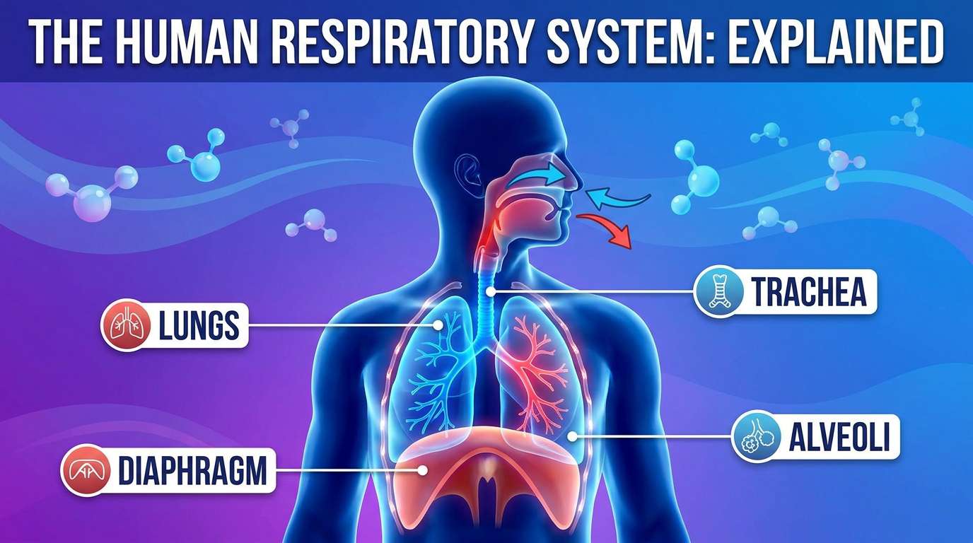
The Human Respiratory System Explained
Introduction
Take a breath right now. Notice how your chest rises slightly, how air moves in through your nose or mouth, travels somewhere deep inside your body, and then flows back out again. You will do this roughly 20,000 times today, almost entirely without conscious thought, and you have been doing it continuously since the moment you were born.
The respiratory system is responsible for one of the most fundamental processes keeping you alive: bringing oxygen into the body and removing carbon dioxide, a waste product of cellular respiration. Without this constant exchange, cells throughout the body would quickly be unable to release the energy they need from food, and within minutes, the brain and other vital organs would begin to fail.
This article explores the respiratory system in depth — its structures, the mechanics of breathing, how gas exchange actually occurs at the cellular level, and the disorders that can affect this vital system.
Why the Body Needs a Respiratory System
Every cell in the human body carries out a process called cellular respiration, in which glucose is broken down to release energy. This process requires a continuous supply of oxygen and produces carbon dioxide as a waste product. If carbon dioxide is allowed to accumulate in the blood, it makes the blood more acidic, which can be dangerous and even fatal if not corrected quickly.
The respiratory system exists to solve two connected problems at once: bringing in the oxygen that cellular respiration requires, and removing the carbon dioxide it produces, keeping the internal environment of the body stable.
The Structures of the Respiratory System
Air travels through a series of connected structures on its way to and from the lungs, each with a specific role in preparing, filtering, or exchanging the air.
Nose and Nasal Cavity
Air normally enters the respiratory system through the nose, where it passes through the nasal cavity. This cavity is lined with fine hairs and a layer of moist mucus, which together filter out dust, pollen, and other particles before they can travel further into the delicate lungs. The nasal cavity also warms and moistens incoming air, protecting the sensitive tissues deeper in the respiratory tract from cold, dry air.
Pharynx
The pharynx, or throat, is a shared passage for both air and food. It connects the nasal cavity and mouth to the next structures in the respiratory tract, while a flap of tissue called the epiglottis ensures that food is directed into the oesophagus (the tube leading to the stomach) rather than into the airway, particularly during swallowing.
Larynx
Below the pharynx lies the larynx, commonly known as the voice box, because it contains the vocal cords. Air passing over the vocal cords causes them to vibrate, producing the sounds used in speech.
Trachea
The trachea, or windpipe, is a tube reinforced with C-shaped rings of cartilage that keep it open even as air pressure changes during breathing. The trachea is lined with cells that produce mucus and with tiny hair-like structures called cilia. Together, the mucus and cilia trap remaining dust and microorganisms and sweep them upward, away from the lungs, to be swallowed or coughed out.
Bronchi and Bronchioles
At its lower end, the trachea divides into two branches called the bronchi (singular: bronchus), one leading to each lung. Inside each lung, the bronchi divide repeatedly into progressively smaller tubes called bronchioles. This branching pattern — a single main tube splitting again and again into finer and finer tubes — is an example of the fractal branching found throughout biological transport systems, allowing air to reach every part of the lung efficiently.
Alveoli
At the very end of the smallest bronchioles are tiny, thin-walled air sacs called alveoli (singular: alveolus). This is where the true purpose of the entire respiratory system is finally achieved: the exchange of oxygen and carbon dioxide between air and blood.
Each lung contains hundreds of millions of alveoli, giving the lungs an astonishingly large internal surface area — often compared to the surface area of a tennis court, despite fitting inside a chest cavity roughly the size of two fists. This enormous surface area is essential for efficient gas exchange to occur quickly enough to meet the body's constant demand for oxygen.
Lungs
The lungs are the two large, spongy organs that house the bronchi, bronchioles, and alveoli. The right lung is slightly larger than the left, which is slightly smaller to accommodate space for the heart. The lungs are protected by the rib cage and sit within an airtight cavity in the chest.
Diaphragm and Intercostal Muscles
The diaphragm is a dome-shaped sheet of muscle located beneath the lungs, separating the chest cavity from the abdominal cavity. Together with the intercostal muscles located between the ribs, the diaphragm is responsible for the physical movements that actually draw air into the lungs and push it back out.
The Mechanics of Breathing
Breathing is not something the lungs do actively by themselves — the lungs have no muscle of their own capable of expanding or contracting. Instead, breathing relies entirely on changes in the volume and pressure of the chest cavity, driven by the diaphragm and intercostal muscles.
Inhalation (Breathing In)
During inhalation:
- The diaphragm contracts and flattens, moving downward.
- The intercostal muscles contract, pulling the ribs upward and outward.
- These movements together increase the volume of the chest cavity.
- This increase in volume causes a decrease in pressure inside the lungs, relative to the air outside the body.
- Air, naturally moving from an area of higher pressure to lower pressure, rushes into the lungs to equalize this difference.
Exhalation (Breathing Out)
During exhalation, the process reverses:
- The diaphragm relaxes and returns to its dome shape, moving upward.
- The intercostal muscles relax, allowing the ribs to move downward and inward.
- These movements decrease the volume of the chest cavity.
- This decrease in volume increases the pressure inside the lungs.
- Air, again moving from higher to lower pressure, is pushed out of the lungs into the surrounding environment.
This entire process, driven by changes in pressure caused by simple, coordinated muscle movements, is what allows air to move in and out of the lungs roughly twelve to twenty times every minute in a resting adult.
Gas Exchange at the Alveoli
The actual exchange of gases between air and blood takes place across the thin walls of the alveoli, which are surrounded by a dense network of capillaries. Several structural features make this exchange remarkably efficient:
- A very large total surface area, provided by the hundreds of millions of alveoli in each lung.
- Extremely thin walls, often just one cell thick, both in the alveoli and the surrounding capillaries, minimizing the distance gases must diffuse.
- A rich capillary network, ensuring a constant, close supply of blood at the exchange surface.
- A moist inner lining, which helps gases dissolve and diffuse more easily across the alveolar wall.
Oxygen, present in much higher concentration in the air within the alveoli than in the blood arriving from the body, diffuses across the alveolar and capillary walls into the blood, where it binds to haemoglobin in red blood cells. At the same time, carbon dioxide, present in higher concentration in the blood than in the air within the alveoli, diffuses in the opposite direction, from the blood into the alveoli, ready to be exhaled.
Comparing Inhaled and Exhaled Air
Although air is a mixture of several gases, its composition changes noticeably after passing through the lungs:
| Gas | Inhaled Air (approx.) | Exhaled Air (approx.) |
|---|---|---|
| Oxygen | 21% | 16% |
| Carbon dioxide | 0.04% | 4% |
| Nitrogen | 78% | 78% (largely unchanged) |
| Water vapour | Variable | Higher (increased by moisture from the lungs) |
Notice that exhaled air still contains a significant amount of oxygen (around 16%) — the body does not extract every trace of oxygen from each breath, which is part of the reason artificial respiration (breathing air directly into another person's lungs) can still provide them with usable oxygen in an emergency.
Cellular Respiration: What Happens to Oxygen After It Enters the Blood
Oxygen absorbed into the blood at the alveoli is transported, bound to haemoglobin within red blood cells, to tissues throughout the body. Once delivered to individual cells, oxygen is used in cellular respiration — a series of chemical reactions that break down glucose to release energy, producing carbon dioxide and water as by-products.
The overall word equation for this process (specifically aerobic respiration, which requires oxygen) is:
Glucose + Oxygen → Carbon dioxide + Water + Energy
The carbon dioxide produced in this process diffuses out of cells into the blood, is transported back to the lungs, and diffuses into the alveoli to be exhaled — completing the connection between breathing and the energy-producing processes happening inside every cell of the body.
Control of Breathing Rate
Breathing rate is normally controlled automatically by a region of the brain called the medulla oblongata, which monitors the level of carbon dioxide in the blood. When carbon dioxide levels rise — for example, during exercise, when cells are respiring more rapidly and producing more carbon dioxide — the medulla oblongata increases the rate and depth of breathing to remove the excess carbon dioxide more quickly and bring in more oxygen.
This is an example of a negative feedback mechanism: a rise in carbon dioxide triggers a response (increased breathing) that acts to reduce carbon dioxide levels back toward normal, maintaining a stable internal environment.
Lung Capacity and Volumes
Not every breath moves the same amount of air, and the lungs are never completely emptied or completely filled during normal breathing. Several terms describe different aspects of lung capacity:
- Tidal volume: The amount of air moved in or out during a single normal breath.
- Vital capacity: The maximum amount of air that can be forcibly exhaled after a maximum inhalation.
- Residual volume: The amount of air that always remains in the lungs, even after the most forceful possible exhalation.
Regular physical exercise can increase vital capacity over time, which is one reason trained athletes are often able to sustain intense physical activity for longer than an untrained individual.
Common Disorders of the Respiratory System
Asthma
Asthma is a condition in which the airways become inflamed and narrowed, often triggered by allergens, cold air, or physical exertion. This narrowing makes it difficult for air to move freely in and out of the lungs, causing wheezing, coughing, and shortness of breath.
Pneumonia
Pneumonia is an infection that causes the alveoli to become inflamed and filled with fluid or pus. Since gas exchange depends on air reaching the alveoli efficiently, fluid-filled alveoli significantly reduce the amount of oxygen that can be absorbed into the blood.
Bronchitis
Bronchitis is inflammation of the lining of the bronchi, often caused by infection or exposure to irritants such as smoke. It leads to excess mucus production and a persistent cough as the body attempts to clear the airway.
Tuberculosis (TB)
Tuberculosis is a serious bacterial infection that primarily affects the lungs, causing damage to lung tissue over time. It remains a significant public health concern in Nigeria and many other countries, spread through airborne droplets from an infected person's cough.
Effects of Smoking on the Respiratory System
Cigarette smoke contains numerous harmful chemicals that damage the respiratory system in several ways: it paralyzes and eventually destroys the cilia lining the trachea and bronchi, allowing dust and pathogens to reach the lungs more easily; it irritates and inflames airway linings, contributing to chronic bronchitis; and it can damage the delicate walls of the alveoli over time, reducing the surface area available for gas exchange — a condition known as emphysema.
How the Respiratory System Connects to Other Body Systems
- Circulatory system: Transports oxygen absorbed in the lungs to body tissues, and carries carbon dioxide back to the lungs for removal.
- Nervous system: The medulla oblongata continuously regulates breathing rate based on blood carbon dioxide levels.
- Muscular system: The diaphragm and intercostal muscles physically drive the mechanics of breathing.
The Respiratory System and WAEC/NECO/JAMB Biology
Key examinable areas of this topic include:
- Labelled diagrams of the respiratory system, from the nasal cavity through to the alveoli.
- The mechanism of inhalation and exhalation, including the specific roles of the diaphragm and intercostal muscles.
- The structural adaptations of the alveoli that make them efficient for gas exchange.
- Comparing the composition of inhaled and exhaled air.
- The word equation for aerobic respiration and its connection to breathing.
- Effects of smoking and common respiratory diseases on lung structure and function.
Common Mistakes Students Make
- Confusing breathing with respiration. Breathing is the physical movement of air in and out of the lungs; respiration is the chemical process inside cells that releases energy from glucose. They are related but not the same thing.
- Describing the diaphragm as an organ that "pumps" the lungs directly. The diaphragm changes chest cavity volume, which changes pressure, which then causes air to move — it does not physically squeeze the lungs.
- Forgetting that exhaled air still contains a significant amount of oxygen, not zero.
- Mislabelling the trachea, bronchi, and bronchioles in diagrams, particularly confusing the order in which air passes through them.
- Omitting cartilage rings when describing the trachea's structure, which are important for explaining why the trachea does not collapse.
Conclusion
The respiratory system is a finely tuned piece of biological engineering, built around a single essential task: bringing air close enough to the blood that oxygen and carbon dioxide can be exchanged efficiently, again and again, tens of thousands of times each day. From the filtering hairs in the nose to the microscopic alveoli deep within the lungs, every structure along the way exists to support this one continuous exchange.
Understanding this system in depth — not just labelling a diagram, but truly grasping how pressure changes drive breathing, and how structure enables gas exchange at the cellular level — gives students a genuine appreciation for one of the quietest, most constant, and most essential processes their own bodies perform every moment of their lives.